Cardiac q‐space trajectory imaging by motion‐compensated tensor‐valued diffusion encoding in human heart in vivo
Diffusion
Diffusion MRI
Diffusion MRI is a non-invasive imaging technique that probes tissue microstructure by measuring the random motion of water molecules.
Introduction
Diffusion MRI is a non-invasive imaging technique that probes tissue microstructure by measuring the random motion of water molecules. Because water mobility is influenced by cellular density, membrane integrity, extracellular space, and tissue organisation, diffusion MRI can provide sensitive information about tissue architecture that is not visible on conventional anatomical imaging.
Diffusion MRI acquisitions are typically performed over a range of b-values, which determine how strongly the images are sensitised to water motion. Lower b-values retain more overall signal and may include contributions from faster-moving components, whereas higher b-values provide greater sensitivity to hindered and restricted diffusion within tissue.
By measuring how signal changes across different b-values, diffusion MRI can derive quantitative parameters such as the apparent diffusion coefficient (ADC), a widely used measure of water mobility within tissues. Diffusion MRI is most commonly acquired using echo planar imaging (EPI), which allows rapid image collection and supports clinically feasible scan times. The choice of b-values and EPI approach is therefore a key part of diffusion MRI study design, as it influences image quality, quantitative analysis, and the physiological meaning of the derived parameters.
Model
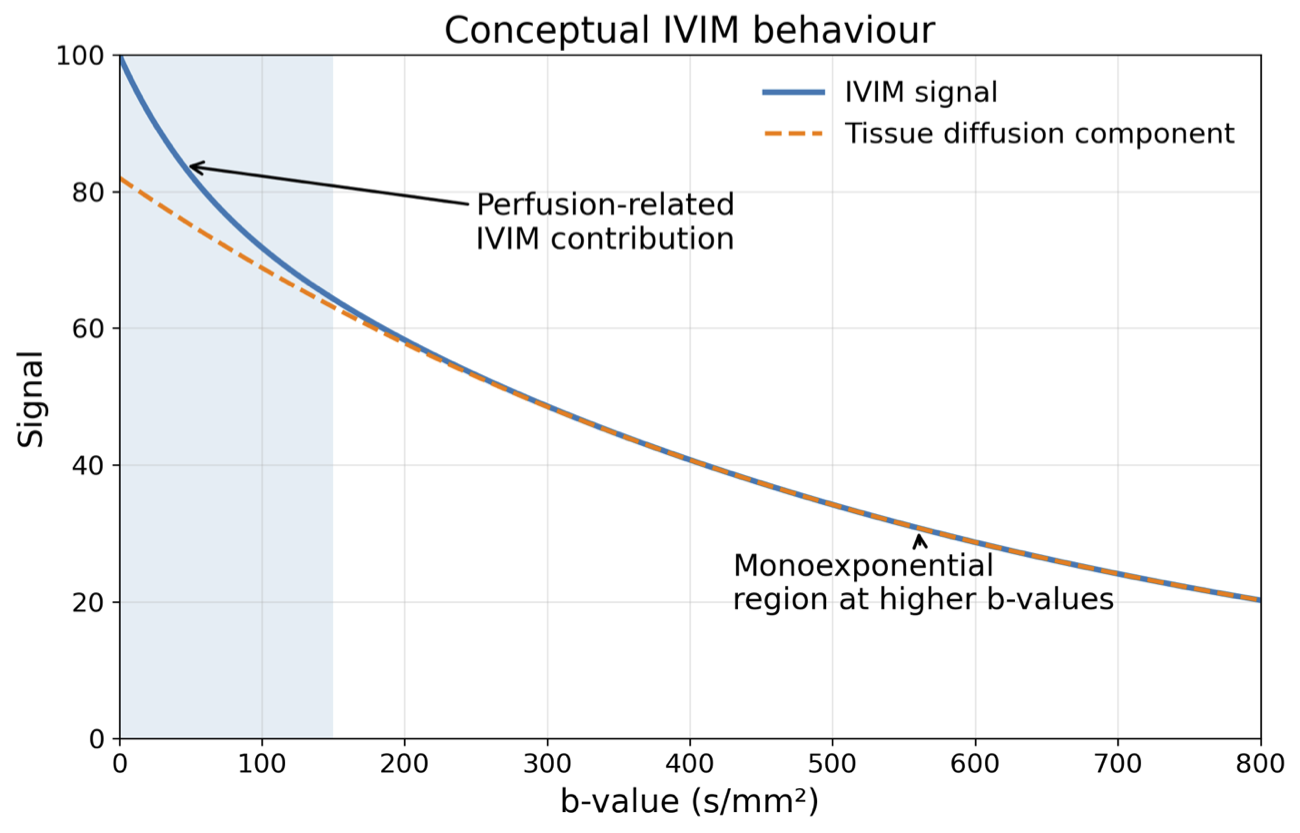
Intravoxel incoherent motion, or IVIM, is a widely used diffusion MRI approach that aims to separate the effects of molecular diffusion from signal changes related to microvascular flow. By acquiring data over a wide range of b-values, IVIM models the signal as a combination of a slower diffusion-related component and a faster component associated with blood moving through the capillary network.
In practical terms, IVIM helps translate diffusion-weighted signal changes into biologically meaningful parameters. The parameter D reflects the true diffusion of water within tissue, while D* represents the pseudo-diffusion component linked to perfusion-related motion, and f represents the perfusion fraction. Together, these parameters can provide insight into both tissue microstructure and microvascular behaviour.
Although mathematically compact, IVIM is powerful because it extends diffusion MRI beyond structural characterisation alone. For this reason, it is widely used in abdominal imaging, oncology, and other applications where both tissue architecture and perfusion-related effects are relevant. Its strength lies in providing quantitative biomarkers without the need for exogenous contrast agents.
Brain
In the brain, diffusion MRI is particularly valuable because tissue microstructure is closely related to function and pathology. Water mobility in brain tissue is influenced by cellular organisation, axonal structure, myelination, oedema, and necrosis, meaning that diffusion measurements can provide important information about disease processes even when structural changes are subtle or absent on conventional MRI.
Diffusion MRI is widely used in neurology and neuro-oncology. It plays a central role in acute stroke imaging, where restricted diffusion can help identify early ischaemic injury. It is also used in brain tumours, inflammatory conditions, and neurodegenerative disease, where changes in diffusion may reflect altered cellularity, tissue disruption, or treatment response. In white matter, diffusion-based approaches can also support assessment of fibre organisation and connectivity.
Oncology
In oncology, diffusion MRI is used to probe tumour cellularity and tissue heterogeneity. Because the mobility of water is often reduced in highly cellular tissue, diffusion measurements can provide a functional readout of tumour microstructure that complements anatomical imaging. This is particularly valuable in cancers, where cellular density, necrosis, stromal content, or treatment-induced change may all influence tissue water diffusion.
Diffusion MRI can help support lesion detection, tissue characterisation, and treatment response assessment. Tumours with low diffusion may reflect densely packed viable tissue, whereas increasing diffusion over time may indicate treatment-related cell death or breakdown of tissue structure. This makes diffusion MRI particularly attractive in longitudinal studies, where changes in diffusion can serve as imaging biomarkers of response before size changes become apparent.
In addition, IVIM can provide complementary information on tumour microvascular status, acting as a contrast-free proxy for perfusion-related behaviour. Together, these approaches allow diffusion MRI to capture both microstructural and vascular aspects of the tumour microenvironment.
In some oncology studies, diffusion MRI is also combined with techniques such as DCE-MRI or OE-MRI to provide complementary information. In this setting, diffusion MRI reflects tissue microstructure, while DCE-MRI characterises vascular delivery and permeability, and OE-MRI provides information related to oxygen-responsive tissue. Together, these methods can offer a more complete picture of the tumour microenvironment.