Bioxydyn offers imaging biomarkers for respiratory diseases such as cystic fibrosis, asthma, pulmonary hypertension, interstitial disease and COPD. We have extensive expertise in quantitative MRI methods for assessing regional lung function, including measurements of ventilation and perfusion.
Bioxydyn has been at the forefront of the development and deployment of quantitative MRI methods for assessing lung disease. Our focus is on the use of proton MRI methods that can be deployed on most hospital MRI scanners without substantial investment in specialist hardware or consumables.
Oxygen-enhanced MRI (OE-MRI) is a non-invasive and non-ionising imaging method that uses oxygen as an MR imaging agent, which is delivered to the patient in the scanner. OE-MRI can be deployed on most hospital scanners.
Lung OE-MRI monitors the delivery of oxygen to patient's lungs. The acquired quantitative imaging biomarkers relate to regional lung ventilation and gas diffusion. Bioxydyn has extensive experience in deploying OE-MRI in diseases including chronic obstructive pulmonary disease (COPD), cystic fibrosis, Primary ciliary dyskinesia (PCD), interstitial lung disease (ILD), lung cancer, and asthma.
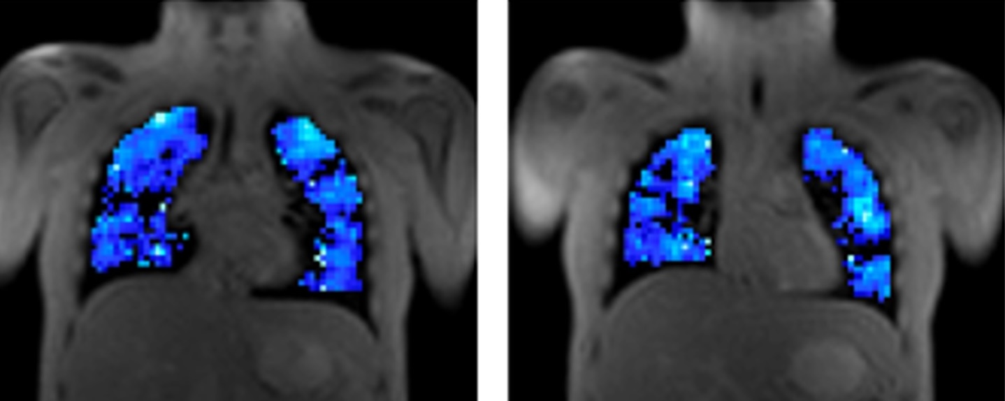
The images show ventilation maps from different coronal slices in an adult cystic fibrosis patient. Blue indicates regions receiving oxygen; black regions indicate defects.
Bioxydyn is also a leader in dynamic contrast-enhanced MRI (DCE-MRI) in the lung, providing information on lung perfusion and other microvascular function, including endothelial permeability and leakage space in diseases such as pulmonary embolism, asthma, pulmonary hypertension, interstitial disease and COPD.
Bioxydyn also provides imaging biomarkers of lung ventilation based on the respiratory motion of the lung over time. These measurements provide ventilation-weighted imaging in a short scan without administration of gases or contrast agents.
In addition to conventional (proton) MRI, Bioxydyn provides services to support the use of hyperpolarised 129Xe MRI in studies of lung function. Working with established academic partners, we offer services to support single and multi-site 129Xe studies investigating ventilation, gas transfer, and pulmonary microstructure.
Bioxydyn offers a range of imaging biomarkers of regional lung function for the assessment of treatment benefit and harm. All can be performed single- or multi-centre using a range of vendor’s equipment. Bioxydyn’s imaging methods bave been validated in multiple studies, including as part of the Innovative Medicines Initiative TRISTAN project, funded by the EU.
In 't Zandt R, Mahmutovic Persson I, Tibiletti M, von Wachenfeldt K, Parker GJM, Olsson LE, TRISTAN Consortium, Contrast enhanced longitudinal changes observed in an experimental bleomycin-induced lung fibrosis rat model by radial DCE-MRI at 9.4T. PLoS One. 2024;19(9):e0310643.
Kim M, Naish JH, Needleman SH, Tibiletti M, Taylor Y, O'Connor JPB, Parker GJM, Feasibility of dynamic T2*-based oxygen-enhanced lung MRI at 3T. Magn Reson Med. 2024 Mar;91(3):972-986.
Needleman SH, Kim M, McClelland JR, Naish JH, Tibiletti M, O'Connor JPB, Parker GJM, Independent component analysis (ICA) applied to dynamic oxygen-enhanced MRI (OE-MRI) for robust functional lung imaging at 3 T. Magn Reson Med. 2024 Mar;91(3):955-971.
Edwards L, Waterton JC, Naish J, Short C, Semple T, Jm Parker G, Tibiletti M, Imaging human lung perfusion with contrast media: A meta-analysis. Eur J Radiol. 2023 Jul;164:110850.
Ohene Y, Harris WJ, Powell E, Wycech NW, Smethers KF, Lasič S, South K, Coutts G, Sharp A, Lawrence CB, Boutin H, Parker GJM, Parkes LM, Dickie BR, Filter exchange imaging with crusher gradient modelling detects increased blood-brain barrier water permeability in response to mild lung infection. Fluids Barriers CNS. 2023 Apr 3;20(1):25.
Tibiletti M, Eaden JA, Naish JH, Hughes PJC, Waterton JC, Heaton MJ, Chaudhuri N, Skeoch S, Bruce IN, Bianchi S, Wild JM, Parker GJM, Imaging biomarkers of lung ventilation in interstitial lung disease from 129Xe and oxygen enhanced 1H MRI. Magn Reson Imaging. 2023 Jan;95:39-49.
Cheriyan J, Roberts A, Roberts C, Graves MJ, Patterson I, Slough RA, Schroyer R, Fernando D, Kumar S, Lee S, Parker GJM, Sarov-Blat L, McEniery C, Middlemiss J, Sprecher D, Janiczek RL, Evaluation of Dynamic Contrast-Enhanced MRI Measures of Lung Congestion and Endothelial Permeability in Heart Failure: A Prospective Method Validation Study. J Magn Reson Imaging. 2022 Aug;56(2):450-461.
Mahmutovic Persson I, von Wachenfeldt K, Waterton JC, Olsson LE, On Behalf Of The Tristan Consortium, Imaging Biomarkers in Animal Models of Drug-Induced Lung Injury: A Systematic Review. J Clin Med. 2020 Dec 30;10(1):.
Jacob J, Alexander D, Baillie JK, Berka R, Bertolli O, Blackwood J, Buchan I, Bloomfield C, Cushnan D, Docherty A, Edey A, Favaro A, Gleeson F, Halling-Brown M, Hare S, Jefferson E, Johnstone A, Kirby M, McStay R, Nair A, Openshaw PJM, Parker G, Reilly G, Robinson G, Roditi G, Rodrigues JCL, Sebire N, Semple MG, Sudlow C, Woznitza N, Joshi I, Using imaging to combat a pandemic: rationale for developing the UK National COVID-19 Chest Imaging Database. Eur Respir J. 2020 Aug;56(2):.
Salem A, Little RA, Latif A, Featherstone AK, Babur M, Peset I, Cheung S, Watson Y, Tessyman V, Mistry H, Ashton G, Behan C, Matthews JC, Asselin MC, Bristow RG, Jackson A, Parker GJM, Faivre-Finn C, Williams KJ, O'Connor JPB, Oxygen-enhanced MRI Is Feasible, Repeatable, and Detects Radiotherapy-induced Change in Hypoxia in Xenograft Models and in Patients with Non-small Cell Lung Cancer. Clin Cancer Res. 2019 Jul 1;25(13):3818-3829.
Skeoch S, Weatherley N, Swift AJ, Oldroyd A, Johns C, Hayton C, Giollo A, Wild JM, Waterton JC, Buch M, Linton K, Bruce IN, Leonard C, Bianchi S, Chaudhuri N, Drug-Induced Interstitial Lung Disease: A Systematic Review. J Clin Med. 2018 Oct 15;7(10):.
Martini K, Gygax CM, Benden C, Morgan AR, Parker GJM, Frauenfelder T, Volumetric dynamic oxygen-enhanced MRI (OE-MRI): comparison with CT Brody score and lung function in cystic fibrosis patients. Eur Radiol. 2018 Oct;28(10):4037-4047.
Alamidi DF, Kindvall SS, Hubbard Cristinacce PL, McGrath DM, Young SS, Naish JH, Waterton JC, Wollmer P, Diaz S, Olsson M, Hockings PD, Lagerstrand KM, Parker GJ, Olsson LE, T1 Relaxation Time in Lungs of Asymptomatic Smokers. PLoS One. 2016;11(3):e0149760.
Zhang WJ, Niven RM, Young SS, Liu YZ, Parker GJ, Naish JH, T1-weighted Dynamic Contrast-enhanced MR Imaging of the Lung in Asthma: Semiquantitative Analysis for the Assessment of Contrast Agent Kinetic Characteristics. Radiology. 2016 Mar;278(3):906-16.
Tiddens HA, Stick SM, Wild JM, Ciet P, Parker GJ, Koch A, Vogel-Claussen J, Respiratory tract exacerbations revisited: ventilation, inflammation, perfusion, and structure (VIPS) monitoring to redefine treatment. Pediatr Pulmonol. 2015 Oct;50 Suppl 40:S57-65.
Zhang WJ, Niven RM, Young SS, Liu YZ, Parker GJ, Naish JH, Dynamic oxygen-enhanced magnetic resonance imaging of the lung in asthma -- initial experience. Eur J Radiol. 2015 Feb;84(2):318-26.
Morgan AR, Parker GJ, Roberts C, Buonaccorsi GA, Maguire NC, Hubbard Cristinacce PL, Singh D, Vestbo J, Bjermer L, Jögi J, Taib Z, Sarv J, Bruijnzeel PL, Olsson LE, Bondesson E, Nihlén U, McGrath DM, Young SS, Waterton JC, Nordenmark LH, Feasibility assessment of using oxygen-enhanced magnetic resonance imaging for evaluating the effect of pharmacological treatment in COPD. Eur J Radiol. 2014 Nov;83(11):2093-101.
Kershaw LE, Naish JH, McGrath DM, Waterton JC, Parker GJ, Measurement of arterial plasma oxygenation in dynamic oxygen-enhanced MRI. Magn Reson Med. 2010 Dec;64(6):1838-42.
Naish JH, Kershaw LE, Buckley DL, Jackson A, Waterton JC, Parker GJ, Modeling of contrast agent kinetics in the lung using T1-weighted dynamic contrast-enhanced MRI. Magn Reson Med. 2009 Jun;61(6):1507-14.